

Dermatologie » Entzündliche Dermatosen » Akne und Rosacea

»

EADV: AI-driven profiling of rosacea severity and markers in clinic and research

Digitale Hände halten leuchtenden AI-Mikrochip – Symbol für Künstliche Intelligenz und Digitalisierung

Quelle: © AiInfo - stock.adobe.com

EADV: AI-driven profiling of rosacea severity and markers in clinic and research

Fachartikel

Dermatologie

Akne und Rosacea

mgo medizin

mgo medizin Redaktion

Verlag

3 MIN

Erschienen in: DermaForum

Objectively assessing rosacea severity is difficult. A study demonstrates an AI approach for standardized, quantitative profiling that goes beyond the mere detection of redness.

Rosacea is a chronic inflammatory skin disease in which objective and reproducible assessment of disease severity remains challenging in both clinical practice and research. In this study we presented an AI-driven imaging approach for standardized, quantitative profiling of rosacea severity beyond visible redness alone.

Using the clinically validated Scarletred® Vision Software Platform in combination with a mobile application and standardized image acquisition via a skin patch, facial images were collected longitudinally from 21 patients (355 images). The analysis is powered by Scarletred® ARORA AI-Agent, a multimodal artificial intelligence engine that coordinates image preprocessing, automated feature extraction, and longitudinal pattern analysis. The AI-Agent automatically quantified multiple rosacea-related morphological markers, including affected facial surface area, erythema intensity, telangiectasia length, and number of papules and pustules.

“With this work, our aim was to move beyond Subjective visual grading and provide dermatologists with an objective, standardized tool for assessing rosacea severity. Our AI-derived morphological biomarkers may support future digital endpoints in clinical trials and enable more personalized treatment strategies.”

Chiara Agostini, MSc., Data Scientist

The AI-based analysis accurately delineated erythematous regions, enabling objective measurement of erythema extension and intensity, while simultaneously detecting and quantifying telangiectatic structures, inflammatory lesions, and estimating the presence and severity of rhinophyma. Longitudinal analysis allowed visualization of erythema intensity dynamics and changes in morphological markers over time, supporting precise monitoring of disease progression and treatment response.
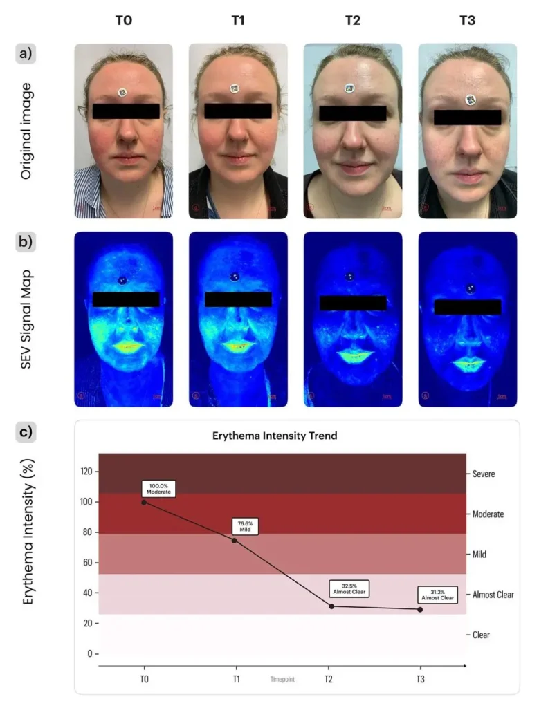
BU: Composite figure showing (a) the original image, (b) the corresponding SEV signal map. (c) the graphical representation of the erythema intensity trend; © Supplementary graphics and patient images, with source attribution (SCARLETRED Holding GmbH, Vienna, Austria, and Department of Dermatology, Universitätsklinikum Tuebingen, Tuebingen, Baden-Wuerttemberg, Germany).
Abb.1: Composite figure showing (a) the original image, (b) the corresponding SEV signal map. (c) the graphical representation of the erythema intensity trend; © Supplementary graphics and patient images, with source attribution (SCARLETRED Holding GmbH, Vienna, Austria, and Department of Dermatology, Universitätsklinikum Tuebingen, Tuebingen, Baden-Wuerttemberg, Germany).

Our results demonstrate the potential of AI-enhanced facial imaging as a standardized, automated, and reproducible framework for rosacea assessment, paving the way toward more personalized and data-driven dermatological care. Our AI-supported framework enables objective, longitudinal rosacea assessment suitable for both routine clinical practice and decentralized monitoring. Unlike conventional ordinal grading systems, it provides continuous, quantitative metrics across multiple morphological dimensions, supporting reproducible digital clinimetry while minimizing inter- and intra-observer variability.
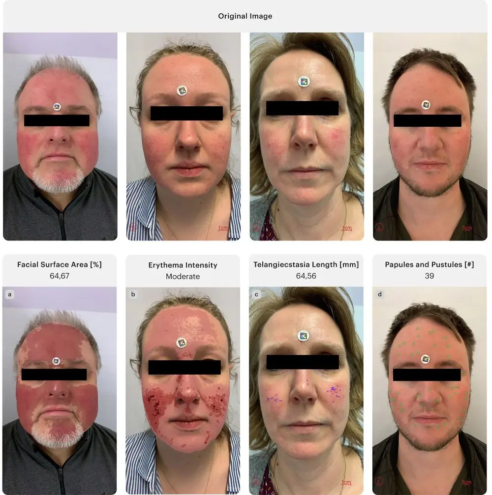
Representative images corresponding to the evaluation of specific clinical parameters. Each image highlights one feature: affected area (a), erythema intensity (b), telangiectasia c), and papules/pustules (d); © Supplementary graphics and patient images, with source attribution (SCARLETRED Holding GmbH, Vienna, Austria, and Department of Dermatology, Universitätsklinikum Tuebingen, Tuebingen, Baden-Wuerttemberg, Germany).

Authors: C. Agostini, M. Molnarova, M. Schaller, and H. Schnidar

Correspondence address:

Chiara Agostini, MSc., Data Scientist

Chiara Agostini, MSc
SCARLETRED Holding GmbH
Wien, Austria

Website: https://www.scarletred.com/

E-Mail: chiara.agostini@scarletred.com


Relevant links to current projects and research related to this work

Schaller, M., Riel, S., Bashur, R., Kurup, N., Schnidar, H., & Fehrenbacher, B. (2022). Ivermectin treatment in rosacea: How novel smartphone technology can support monitoring rosacea-associated signs and symptoms. Dermatologic Therapy, 35(11), Artikel e15869. https://doi.org/10.1111/dth.15869

Ranjan, R., Partl, R., Erhart, R., Kurup, N., & Schnidar, H. (2021). The mathematics of erythema: Development of machine learning models for artificial intelligence assisted measurement and severity scoring of radiation induced dermatitis. Computers in Biology and Medicine, 139, Artikel 104952. https://doi.org/10.1016/j.compbiomed.2021.104952

Partl, R., Jonko, B., Schnidar, S., Schöllhammer, M., Bauer, M., Singh, S., Simeckova, J., Wiesner, K., Neubauer, A., &

Schnidar, H. (2017). 128 SHADES OF RED: Objective remote assessment of radiation dermatitis by augmented digital skin imaging. Studies in Health Technology and Informatics, 236, 363–374: https://pubmed.ncbi.nlm.nih.gov/28508819/


Weitere Beiträge zu diesem Thema

Person mit Kapuzenpulli hebt kraftvoll die Arme vor Sonnenaufgang als Symbol fuer Resilienz bei Stress Leistungsabfall und Hautentzuendungen

Stress, Leistungsabfall und Hautentzündungen

News

Sport ist ein starker „Schlafmacher“: Die DGSM betont zum Aktionstag „Erholsamer Schlaf“, dass regelmäßige Bewegung die Schlafqualität verbessert – und damit auch Regeneration und Leistungsfähigkeit. Schlafmangel erhöht Stress (u. a. Cortisol) und kann Entzündungsprozesse sowie chronische Dermatosen triggern. Wichtig ist das Timing: moderates Training ca. 1 h vor dem Schlafen, intensive Einheiten eher mit längerem Abstand.

Dermatologie

Allgemeine Dermatologie

Prävention und Vorsorge

Beitrag lesen
Surreale Collage mit Mensch und Megafon Kopf aus dem mehrfach das Wort FAKE schallt als Symbol fuer digitale Desinformation zur Hautkrebspraevention

Hautkrebsprävention in Zeiten digitaler Desinformation

News

Hautkrebsprävention trifft Social‑Media‑Mythen: Eine Videoanalyse der ADP zeigt, wie Trends („Sonnencreme macht Krebs“, „kein Sonnenbrand = kein Schaden“, „Öle statt LSF“) im Hochsommer riskantes Verhalten fördern. Wichtig für die Praxis: evidenzbasiert, empathisch gegensteuern und den Dreiklang „Meiden, Kleiden, Cremen“ klar kommunizieren – UV‑Schutz als Alltagsroutine.

Dermatologie

Allgemeine Dermatologie

Prävention und Vorsorge

Beitrag lesen
Lupe vergroessert ein Dokument mit Checkliste auf blauem Hintergrund als Symbol fuer die Evidenzpruefung von Kochsalzinjektionen bei kortikosteroidinduzierter Hautatrophie

Kochsalzinjektionen bei kortikosteroidinduzierter Hautatrophie

News

Kochsalzinjektionen könnten kortikosteroidinduzierte Hautatrophie verbessern: Eine systematische Übersicht (16 Artikel, 41 Patient:innen, 48 Läsionen) berichtet nach intraläsionalen 0,9%-NaCl‑Injektionen in 83,3 % vollständige Resolution und bei Hypopigmentierung in 90,9 % Besserung. UAW waren selten. Die Methode gilt als minimalinvasiv und günstig, die Evidenz bleibt heterogen.

Dermatologie

Allgemeine Dermatologie

Versorgung, Forschung und Leitlinien

Beitrag lesen